
Introduction: Building the Foundation for Implant Success
The long-term success of dental implants, the pinnacle of modern tooth replacement, is not merely dependent on the implant itself but is fundamentally anchored to the quality and volume of the patient’s alveolar bone. Following tooth extraction, the jawbone undergoes a natural process of resorption, leading to a loss of both height and width. This often leaves a deficient bony architecture that is inadequate for ideal implant placement. This is where the science of bone grafting becomes the clinician’s most powerful tool.
Bone grafting in dentistry is no longer a niche procedure but a standard of care for predictable, esthetic, and functional implant outcomes. It is the essential step in site development, transforming a compromised ridge into a solid foundation. This definitive guide provides a deep dive into the modern landscape of bone grafting materials for dental implants. We will explore the different classes of materials, their biological mechanisms, and their specific applications in key procedures like socket preservation, ridge augmentation, and the sinus lift, equipping clinicians with the knowledge to generate confident, evidence-based material selections.
The Rationale for Grafting: Why the Jawbone Needs Reinforcement
Understanding the biological imperative for grafting is the first step. When a tooth is lost, the alveolar bone, which exists solely to support that tooth, loses its primary stimulus. This initiates a cascade of remodeling that results in significant bone loss, with the most rapid resorption occurring within the first year.
Without intervention, this can lead to both functional and esthetic compromises for future implant placement. Bone grafting addresses this challenge through three primary clinical procedures:
Socket Preservation: Preventing Collapse After Extraction
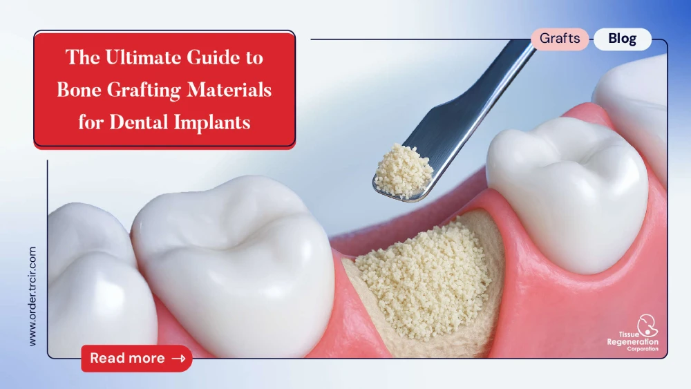
This is the most proactive form of bone grafting. Immediately following a tooth extraction, a graft material is placed into the empty socket. The goal is not to fill a void but to counteract the natural resorption process. Effective socket preservation materials provide a scaffold that supports the surrounding soft tissue and encourages new bone to fill the space, preserving the natural contours of the alveolar ridge for a future implant.
Ridge Augmentation: Rebuilding Lost Bone Volume
When a tooth has been missing for some time, significant bone loss may have already occurred. Ridge augmentation is a surgical procedure to rebuild the height and/or width of the alveolar ridge. This often involves using particulate grafts, sometimes in block form, in conjunction with a barrier membrane in a technique known as Guided Bone Regeneration (GBR). The choice of ridge augmentation materials is critical as they must not only stimulate new bone but also often provide structural support during the healing phase.
Sinus Lift (Sinus Augmentation): Creating Vertical Height in the Maxilla
The maxillary posterior region often presents a dual challenge: insufficient bone height due to both ridge resorption and the natural anatomy of the maxillary sinus. A sinus lift procedure involves elevating the floor of the sinus and placing a bone graft underneath it. This creates the necessary vertical bone height to securely place implants. Selecting the best bone graft for a sinus lift is crucial, as the material must predictably convert to vital bone in this unique anatomical space.

The Biological Principles: What Defines an Ideal Grafting Material?
The efficacy of any bone graft material is judged by its ability to facilitate new bone formation. This is governed by three key biological principles:
- Osteoconduction: The material acts as a biocompatible scaffold or “trellis.” It does not actively contribute to bone formation but provides the physical framework upon which the host’s own bone-forming cells can migrate, attach, and proliferate. This is the most fundamental property of most commercially available graft materials.
- Osteoinduction: The material contains biological agents, such as Bone Morphogenetic Proteins (BMPs), that actively signal and recruit the patient’s undifferentiated stem cells to the site and stimulate them to become osteoblasts (bone-forming cells).
- Osteogenesis: The material itself contains living, viable cells (osteoblasts) that can directly synthesize new bone. This property is exclusive to autografts.
Beyond these principles, factors like a material’s resorption profile, handling characteristics, and structural integrity are also critical to its clinical success.
A Clinician’s Catalog of Bone Grafting Materials
The modern dentist has access to a wide array of grafting materials, each with a unique profile of advantages and limitations.
1. Autografts: The Patient’s Own Gold Standard
An autograft is bone harvested from the patient, either from an intraoral source (like the chin or ramus) or an extraoral source (like the iliac crest).
- Advantages: It is the only material that is fully osteogenic, osteoinductive, and osteoconductive. As it is the patient’s own tissue, there is no risk of disease transmission or immunologic rejection. It remains the biological gold standard.
- Disadvantages: The primary drawback is the need for a second surgical site, which introduces donor site morbidity (pain, swelling, potential nerve damage). The available volume, especially from intraoral sites, is limited.
2. Allografts: The Versatile Human Donor Option
Allografts are sourced from meticulously screened human donors and processed by accredited tissue banks. They are the most widely used graft type in dentistry.
- Description: They are available primarily as a dental allograft powder in two main forms:
- Freeze-Dried Bone Allograft (FDBA): This is a mineralized graft. The processing preserves the mineral content, making it an excellent osteoconductive scaffold with a slow resorption rate, ideal for maintaining space.
- Demineralized Freeze-Dried Bone Allograft (DFDBA): An acid is used to remove the mineral content, exposing the underlying collagen matrix and growth factors (BMPs). This makes DFDBA osteoinductive in addition to being osteoconductive.
- Advantages: Allografts eliminate donor site morbidity, are available in unlimited quantities, and have a proven track record of safety and efficacy. They offer excellent handling and are a workhorse material for socket preservation and ridge augmentation.
- Disadvantages: Processing significantly reduces or eliminates any osteogenic potential. While the risk of disease transmission is statistically infinitesimal (less than 1 in a million), it remains a theoretical consideration.
3. Xenografts: The Cross-Species Contender
Xenografts are derived from a non-human species, most commonly bovine (cow) or porcine (pig). The tissue undergoes a rigorous process to remove all organic components, leaving only the mineral scaffold.
- Advantages: The resulting anorganic bone matrix has a crystalline structure similar to human bone, making it an exceptional osteoconductive scaffold. Its most notable feature is its extremely slow resorption rate, allowing it to act as a long-term volume maintainer, which is particularly useful in large defects or sinus lifts.
- Disadvantages: Xenografts are strictly osteoconductive and have no inductive potential. There can be cultural or religious objections from patients, and a theoretical risk of cross-species disease transmission, though no cases have been documented with modern processing.
4. Alloplasts (Synthetics): The Engineered Alternative
Alloplasts are synthetic, lab-created materials that contain no human or animal tissue. They are typically composed of calcium phosphate-based minerals.
- Description: Common types include Hydroxyapatite (HA), Beta-Tricalcium Phosphate (β-TCP), and bioactive glasses. They are available in various porosities and resorption profiles.
- Advantages: They carry zero risk of disease transmission and are available in infinite supply. Their resorption rates can be engineered, with β-TCP resorbing faster and HA lasting much longer.
- Disadvantages: They are purely osteoconductive. Some forms can be brittle or difficult to handle. The quality and clinical performance can vary significantly between manufacturers.

Matching the Material to the Mission: A Clinical Decision Framework
There is no single “best” material. The optimal choice depends entirely on the clinical objective.
| Procedure | Primary Goal | Popular Material Choices | Rationale |
| Socket Preservation | Prevent ridge collapse, maintain volume | FDBA Allograft, Xenograft | These materials have slow resorption rates, acting as excellent space maintainers while new bone grows in. |
| Sinus Lift | Create and maintain space for new bone | Xenograft, Allograft, or a composite mix | The very slow resorption of xenograft is a major advantage here, as it prevents graft collapse from sinus pressure. |
| Ridge Augmentation (GBR) | Rebuild horizontal/vertical bone | DFDBA Allograft, Autograft/Allograft mix | Requires an inductive signal. DFDBA’s osteoinductivity is valuable. Mixing with autograft provides an osteogenic boost. |
The Critical Role of Barrier Membranes
Successful bone grafting, especially for ridge augmentation, is not just about the material—it’s about controlling the healing environment. Guided Bone Regeneration (GBR) is a technique that uses a barrier membrane to cover the graft site. This membrane prevents the fast-growing soft tissue cells from invading the graft space, allowing the slower-growing bone cells the time and space they need to populate the scaffold. The utilization of a membrane is considered the standard of care for predictable ridge augmentation.
Conclusion: An Evidence-Based Approach to Predictable Implant Success
The evolution of bone grafting materials for dental implants has fundamentally changed the scope of implant dentistry, allowing clinicians to predictably restore function and esthetics even in severely compromised sites. The choice between an autograft, allograft, xenograft, or alloplast is not arbitrary; it is a clinical decision based on a thorough understanding of the material’s biological properties and the specific demands of the surgical site.
While the autograft remains the biological gold standard, the safety, versatility, and unlimited supply of materials like dental allograft powder have made them the cornerstone of modern implant site development. By mastering the principles of bone biology and thoughtfully selecting the appropriate material for each unique clinical challenge, clinicians can create a solid and enduring foundation, ensuring the highest probability of long-term implant success.
Frequently Asked Questions (FAQ)
1. How long does a dental bone graft take to heal before an implant can be placed?
Healing time varies depending on the size of the defect and the material used, but a general timeframe is 4 to 6 months. The clinician will confirm sufficient bone maturation, often with a new CBCT scan, before proceeding with implant placement.
2. What’s the difference between FDBA and DFDBA allografts?
FDBA (Freeze-Dried Bone Allograft) is mineralized and is primarily osteoconductive, acting as a great scaffold. DFDBA (Demineralized Freeze-Dried Bone Allograft) has its mineral component removed, exposing growth factors that generate it osteoinductive, meaning it can signal new bone formation.
3. Can the body reject a dental bone graft?
True rejection is extremely rare, especially with allografts, xenografts, and alloplasts, as they are processed to be non-immunogenic. The most common cause of graft failure is not rejection but rather infection or lack of blood supply to the site.
4. Why is bovine (cow) bone used for human dental grafts?
Bovine bone, after being processed to remove all organic material, leaves a pure mineral scaffold (anorganic bone matrix). This scaffold has a porosity and structure that is remarkably similar to human bone, making it an excellent and highly predictable osteoconductive material.
5. Do I always need a bone graft after a tooth extraction?
Not always, but it is often recommended, especially if a dental implant is planned for the future. Placing a graft for socket preservation is a minimally invasive way to prevent significant bone loss, which can simplify and improve the outcome of a future implant procedure.
Scientific Sources
- Review of Grafting Materials:
- El-Bialy, T., & El-Bialy, A. (2015). Bone regeneration in dentistry: A review of the literature. The Journal of Contemporary Dental Practice, 16(5), 414-421.
- Link: https://www.jaypeejournals.com/ecom/html/jp/JCDP/JCDP_2015_05_16_5_414_421.pdf
- Guided Bone Regeneration (GBR) Principles:
- Elgali, I., Omar, O., Dahlin, C., & Thomsen, P. (2017). Guided bone regeneration: A systematic review of the effect of barrier membranes on bone regeneration. Journal of Clinical Periodontology, 44(S18), O1-O15.
- Link: https://www.ncbi.nlm.nih.gov/pmc/articles/PMC5643444/
- Socket Preservation Efficacy:
- MacBeth, N., Trullenque-Eriksson, A., & Donos, N. (2017). Hard and soft tissue changes following alveolar ridge preservation: a systematic review. Clinical Oral Implants Research, 28(8), 982–1004.
- Link: https://onlinelibrary.wiley.com/doi/10.1111/clr.12911
- Allograft vs. Xenograft in Sinus Augmentation:
- Corbella, S., Taschieri, S., & Del Fabbro, M. (2018). Long-term outcomes for the management of the edentulous posterior maxilla: a systematic review of allograft vs. xenograft. International Journal of Implant Dentistry, 4(1), 2.
- Link: https://www.ncbi.nlm.nih.gov/pmc/articles/PMC5787037/