
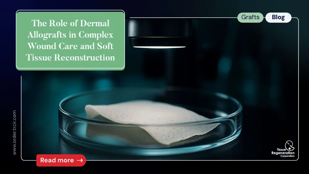
The management of complex wounds and significant soft tissue defects presents a formidable challenge in reconstructive surgery. These defects can arise from trauma, extensive tumor resection, burns, chronic ulcers (e.g., diabetic, venous, pressure), and revision surgeries. Traditional methods, such as primary closure, skin grafting, and local or free flaps, are foundational but may be limited by donor site morbidity, insufficient tissue availability, or patient comorbidities [1]. In this context, acellular dermal allografts (ADAs) have emerged as a pivotal adjunct and, in some cases, a primary solution. Derived from donated human skin that undergoes rigorous processing to remove all cellular components (epidermis and dermal cells), these grafts provide a biocompatible, sterile, and structurally intact dermal scaffold. This article explores the biological principles, clinical applications, and evidence-based role of dermal allografts in modern complex wound care and soft tissue reconstruction.
Biological Principles and Processing of Dermal Allografts
The efficacy of an ADA lies in its ability to serve as a biological scaffold that supports and guides host tissue regeneration. The ideal dermal allograft retains the original three-dimensional architecture of the dermis, including collagen and elastin fibers, as well as a preserved basement membrane complex on one side [2]. This structure confers several key biological advantages:
- Biocompatibility: The removal of cellular components (keratinocytes, fibroblasts, endothelial cells) and major histocompatibility complex (MHC) antigens minimizes the risk of an inflammatory or immune-mediated rejection response from the host [3]. This allows the graft to integrate rather than being rejected.
- Regenerative Properties (Scaffolding): The intact extracellular matrix (ECM) provides a natural, porous scaffold that facilitates host fibroblast and endothelial cell migration. This process, known as re-population and re-vascularization, is essential for transforming the acellular graft into living, functional host tissue [4].
- Preservation of the Basement Membrane: The basement membrane complex, rich in laminin and type IV collagen, provides a critical interface for epithelial cell migration from the wound edges, promoting more rapid and stable re-epithelialization when the graft is placed dermis-side down [5].
Processing is a critical determinant of an ADA’s final properties. While methods vary by manufacturer, they generally involve:
- Decellularization: Using a combination of detergents, enzymatic digestion, and osmotic solutions to remove all cellular material.
- Sterilization: Typically through low-dose gamma irradiation or proprietary chemical sterilization to ensure safety without excessively damaging the collagen structure.
- Preservation: Most commonly freeze-drying (lyophilization) or sterile liquid preservation, which affects the graft’s handling characteristics, rehydration time, and storage requirements [6].
Clinical Applications in Complex Wound Care
Dermal allografts have become indispensable in managing a spectrum of difficult-to-heal wounds.
- Diabetic Foot Ulcers (DFUs): DFUs are a leading cause of lower-limb amputations. When standard care (debridement, offloading, moist wound care) fails, ADAs can be applied to provide a robust scaffold over exposed tendon, bone, or joint capsule, facilitating granulation tissue formation and preparing the wound bed for definitive closure [7].
- Venous Leg Ulcers (VLUs): For large, chronic VLUs refractory to compression therapy, ADAs can help break the cycle of chronic inflammation and stalled healing by providing a healthy ECM, promoting angiogenesis, and accelerating wound closure [8].
- Acute and Chronic Non-healing Wounds: In cases of traumatic wounds with significant tissue loss (degloving injuries), surgical wounds with dehiscence, or pressure ulcers, ADAs serve as a bridge to definitive closure. They provide immediate biological coverage, reduce fluid and protein loss, protect vital underlying structures, and improve the quality of the final reconstructed tissue [9].
Role in Soft Tissue Reconstruction
Beyond wound care, dermal allografts play a significant role in various reconstructive procedures.
- Breast Reconstruction: Following mastectomy, ADAs are widely used to create an inferolateral sling or pocket to support and control the position of a tissue expander or permanent implant. This technique provides better definition of the inframammary fold, reduces implant visibility and palpability (rippling), and may lower the rates of capsular contracture [11].
- Abdominal Wall Reconstruction (Hernia Repair): In complex ventral hernia repairs, particularly in contaminated fields where synthetic mesh is contraindicated, ADAs can be used as a biologic mesh to reinforce the repair. They provide a scaffold for native tissue ingrowth, gradually remodeling into functional host tissue and reducing the risk of chronic infection or fistula formation associated with permanent synthetic materials [12].
- Head and Neck Reconstruction: ADAs are used for skull base repair to prevent cerebrospinal fluid (CSF) leaks, for orbital floor reconstruction, and to augment soft tissue volume in contour defects. Their pliability and low immunogenicity make them well-suited for these delicate anatomical regions [13].
- Tendon and Ligament Augmentation: In orthopedic surgery, ADAs can be used to reinforce repairs of large rotator cuff tears or Achilles tendon ruptures, providing additional mechanical strength and a scaffold for tissue regeneration [14].
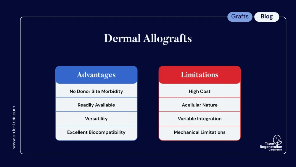
Advantages and Limitations of Dermal Allografts
(Advantages):
- No Donor Site Morbidity: Eliminates pain, scarring, and potential complications associated with harvesting autologous tissue.
- Readily Available: Off-the-shelf availability in various sizes and thicknesses, reducing operative time.
- Versatility: Applicable across a wide range of surgical specialties and clinical scenarios.
- Excellent Biocompatibility: Low rates of rejection and infection when used appropriately.
(Limitations):
- Cost: Dermal allografts are significantly more expensive than traditional wound care dressings or autografts.
- Acellular Nature: Lacking a native blood supply, they are susceptible to failure in poorly vascularized or infected wound beds. Meticulous wound bed preparation is paramount.
- Variable Integration: The time to full vascularization can be unpredictable and is typically slower than that of an autograft.
- Mechanical Properties: While strong, their initial mechanical strength is less than that of synthetic mesh or native fascia, and they undergo a period of remodeling where strength can temporarily decrease before increasing again with host cell repopulation [15].
Conclusion: The Expanding Role of Dermal Allografts
Acellular dermal allografts have revolutionized the approach to complex wound care and soft tissue reconstruction. By providing a structurally intact, biocompatible, and readily available scaffold, they facilitate host tissue integration and regeneration in clinical scenarios where traditional methods fall short. From closing chronic diabetic foot ulcers to augmenting complex breast and abdominal wall reconstructions, ADAs have demonstrated clear clinical utility. The key to successful outcomes lies in appropriate patient and wound selection, meticulous surgical technique, and a thorough understanding of the biological principles governing graft incorporation. As processing technologies continue to advance and clinical evidence expands, the role of dermal allografts is poised to grow further, solidifying their status as an essential tool in the reconstructive surgeon’s armamentarium.
References
- Janis, J. E., & Harrison, B. (2014). Wound healing: part I. Basic science. Plastic and Reconstructive Surgery, 133(2), 199e-207e.
- Wainwright, D. J. (1995). Use of an acellular allograft dermal matrix (AlloDerm) in the management of full-thickness burns. Burns, 21(4), 243-248.
- Hodde, J. P. (2002). Extracellular matrix-based scaffolds for tissue-engineered constructs. Expert Opinion on Biological Therapy, 2(6), 635-646.
- Badylak, S. F., Freytes, D. O., & Gilbert, T. W. (2009). Extracellular matrix as a biological scaffold material: Structure and function. Acta Biomaterialia, 5(1), 1-13.
- Jones, I., Currie, L., & Martin, R. (2002). A guide to biological skin substitutes. British Journal of Plastic Surgery, 55(3), 185-193.
- Cheng, H. W., Feng, S. W., Wu, M. H., & Chen, C. H. (2017). The effects of different preservation methods on the structural and biocompatibility properties of acellular dermal matrix. Journal of Medical and Biological Engineering, 37(3), 336-345.
- Cazzell, S., Vayser, D., Pham, H., Vande Zandsen, D., & Walters, J. (2017). A randomized clinical trial of a human acellular dermal matrix for the closure of diabetic foot ulcers in the wound, ostomy, and continence clinic setting. Ostomy Wound Management, 63(10), 18-29.
- Serena, T. E., Snyder, R. J., & Kirsner, R. S. (2016). Use of a dehydrated human amnion/chorion membrane allograft for the treatment of venous leg ulcers. Wound Repair and Regeneration, 24(5), 878-883.
- Demirkol, M., & Uysal, A. (2010). Clinical experience with acellular dermal matrix in reconstructive surgery. Journal of Plastic, Reconstructive & Aesthetic Surgery, 63(1), 101-108.
- Spear, S. L., & Seruya, M. (2010). Acellular dermal matrix for the treatment and prevention of implant-associated breast deformities. Plastic and Reconstructive Surgery, 126(6), 1773-1781.
- Breuing, K. H., & Warren, S. M. (2005). Immediate bilateral breast reconstruction with implants and AlloDerm. Annals of Plastic Surgery, 55(3), 232-239.
- Hiles, M., & Record, M. C. (2008). The use of allograft dermal matrix in the treatment of complex abdominal wall defects. American Journal of Surgery, 195(1), 7-13.
- Patel, P. N., & Shah, K. S. (2013). Acellular dermal matrix in head and neck reconstruction. Current Opinion in Otolaryngology & Head and Neck Surgery, 21(4), 366-371.
- Barber, F. A., & Burns, J. P. (2006). The use of a xenograft-derived acellular dermal matrix for rotator cuff repair. Arthroscopy, 22(8), 901-905.
- Debare, T., Debeer, P., & Lecouvet, F. E. (2012). The mechanical properties of acellular dermal matrix: a systematic review of the literature. Journal of Plastic, Reconstructive & Aesthetic Surgery, 65(9), 1143-1151.