
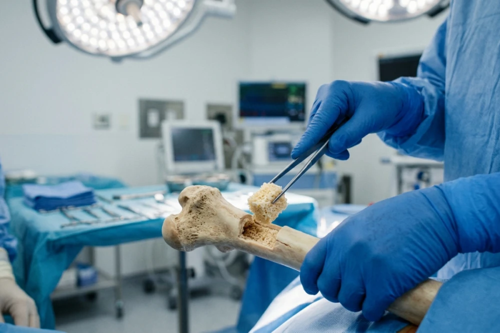
In the rapidly evolving landscape of orthopedic surgery, managing bone defects remains one of the most significant challenges for surgeons worldwide. Whether caused by high-energy trauma, benign bone tumors, or the sequelae of revision arthroplasty, the need to restore skeletal integrity is paramount. This is where the bone void filler steps in as a critical component of modern regenerative medicine.
At TRC (order.trcir.com), we understand that for medical professionals, selecting the right graft material is not just about filling a space; it is about initiating a biological process that leads to structural restoration and patient recovery. In this detailed guide, we will explore the science, types, and clinical applications of bone void fillers, focusing on the latest advancements in allograft bone and tissue regeneration.
What is a Bone Void Filler?
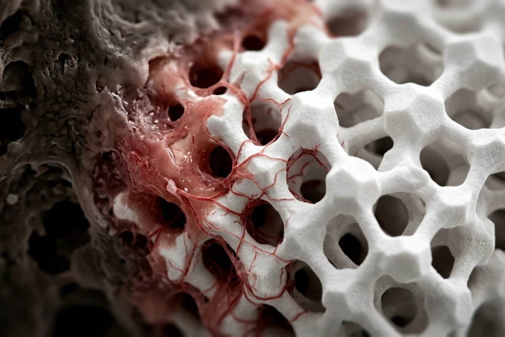
A bone void filler is a sterile, biocompatible material used to fill gaps or voids in the skeletal system. These voids may be the result of surgery, trauma, or degenerative diseases. Unlike simple bone cement, which acts primarily as a grout, modern void fillers are designed to participate actively in the healing process.
The primary function of these materials is to provide a scaffold that supports the ingrowth of new host bone. This process is essential because, in critical-sized defects, the body’s natural regenerative capacity is often insufficient to bridge the gap without architectural support.
The Biological Triad: How It Works
To understand the efficacy of any bone void filler, one must look at the three pillars of bone regeneration. When you browse the catalog at order.trcir.com, you are looking for products that satisfy one or more of these criteria:
Osteoconduction
This is the most fundamental property. An osteoconductive material serves as a scaffold or a trellis. It allows vascular capillaries and bone-forming cells (osteoblasts) to migrate from the host bed into the graft site. Most synthetic fillers and structural allografts excel in this area.
Osteoinduction
Osteoinduction refers to the ability of the material to recruit immature stem cells and stimulate them to differentiate into pre-osteoblasts. This is largely driven by growth factors such as Bone Morphogenetic Proteins (BMPs). Demineralized Bone Matrix (DBM) is a prime example of an osteoinductive bone void filler.
Osteogenesis
This occurs when the graft material actually contains vital, living bone cells that contribute to bone formation. This is typically found in autografts (bone taken from the patient) or specialized cellular allografts.
Classification of Bone Void Fillers
For orthopedic surgeons and procurement managers, distinguishing between the types of fillers is vital for matching the product to the clinical indication.
- Autografts (The Gold Standard)
Autologous bone is harvested from the patient’s own body, usually the iliac crest. While it possesses all three properties (osteoconduction, induction, and genesis), it comes with significant downsides: donor site morbidity, increased surgical time, and limited quantity.
- Allografts
This category is the cornerstone of TRC’s offerings. Allograft bone is derived from human donors and processed under strict sterile conditions.
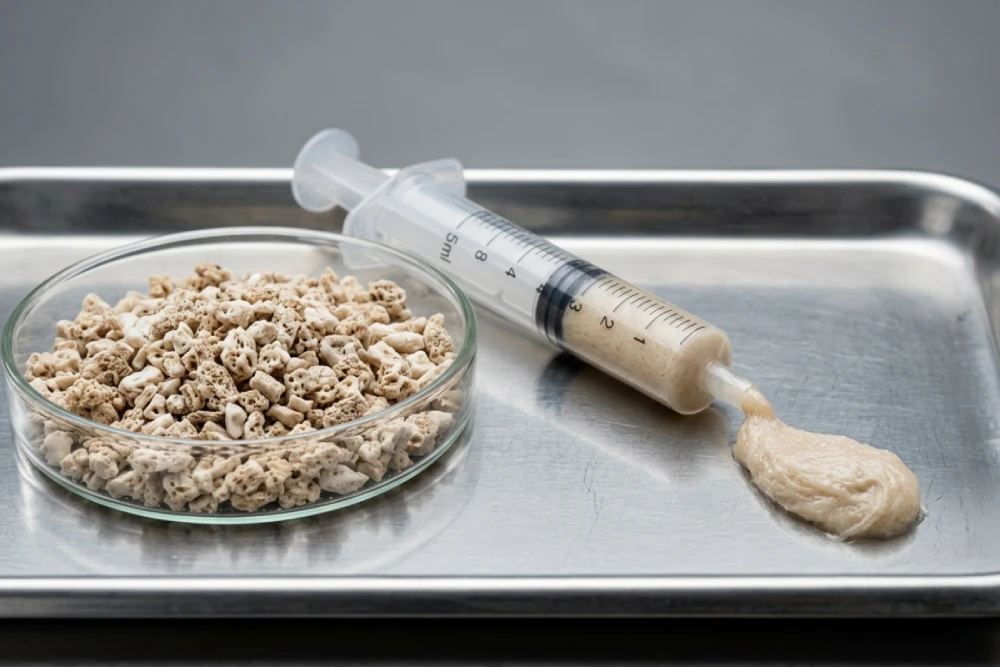
- Mineralized Allografts: Such as cancellous chips or cortical struts, providing excellent structural support and osteoconduction.
- Demineralized Bone Matrix (DBM): The mineral phase is removed to expose the underlying collagen and growth factors, making it highly osteoinductive and this type is provided in different forms of products.
- Advantages: Eliminates donor site pain, available in large quantities, and comes in various forms (chips, putty, gel, strips and etc).
- Synthetic Bone Substitutes
These are man-made materials designed to mimic the mineral composition of bone.
- Ceramics: Hydroxyapatite (HA) and Tricalcium Phosphate (TCP). These are osteoconductive and have a slow resorption rate.
- Calcium Sulfate: Resorbs quickly and is often used as a carrier for antibiotics.
- Bioactive Glass: Bonds directly to bone and stimulates osteoblast activity.
Clinical Applications in Orthopedic Surgery
The versatility of a high-quality bone void filler allows it to be utilized across a wide spectrum of orthopedic procedures.
Trauma and Fracture Management
In comminuted fractures where bone loss is significant (e.g., tibial plateau fractures), the structural integrity of the reduction can be compromised. Packing the defect with cancellous allograft chips helps maintain articular congruity and speeds up union.
Spine Fusion Surgery
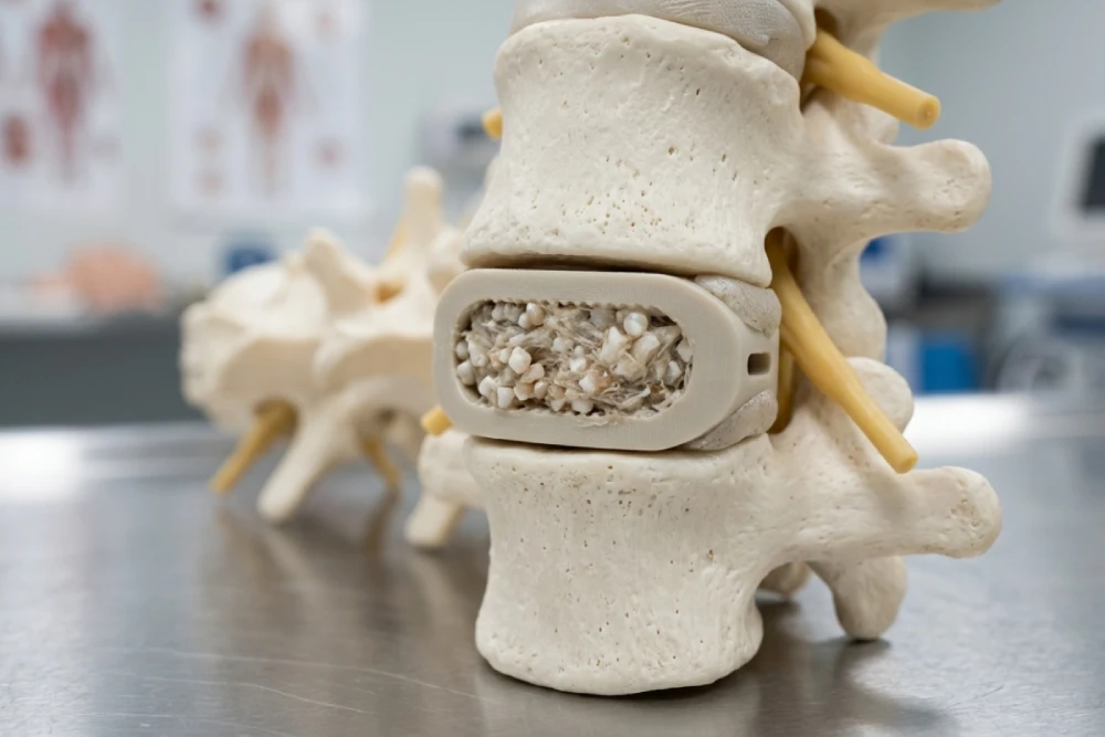
Spinal fusions are among the most common consumers of bone graft materials. To achieve a solid interbody fusion, surgeons often utilize a cage packed with a bone void filler like DBM putty or high-quality allograft. This promotes the formation of a bony bridge between vertebrae, ensuring long-term stability.
Tumor Resection
After the curettage of benign bone cysts or tumors, a large cavity remains. Leaving this empty increases the risk of pathologic fracture. Filling this cavity with a reliable orthopedic implant material restores mechanical strength immediately and encourages remodeling over time.
Revision Arthroplasty
When replacing a failed hip or knee implant, surgeons often encounter massive osteolysis (bone loss). Impacted bone grafting using morselized allograft is a proven technique to restore bone stock before implanting the new prosthesis.
Why Choose Allografts from TRC?
When sourcing products for hospitals and clinics, quality assurance is non-negotiable. At order.trcir.com, our focus on providing top-tier tissue bank products ensures that surgeons can operate with confidence.
- Safety Profile: Our allografts undergo rigorous screening and sterilization processes (such as gamma irradiation or proprietary chemical cleansing) to virtually eliminate the risk of disease transmission while preserving biological activity.
- Structural Integrity: We understand that a bone void filler often needs to bear a load. Our structural allografts are tested to ensure they meet the biomechanical demands of the human body.
- Ease of Handling: Whether you need flowable pastes for minimally invasive injections or compressible sponges for irregular voids, modern allografts are designed for intraoperative efficiency.
Emerging Trends: The Future of Bone Filling
The field is moving towards “smart” biomaterials. We are seeing a rise in composite grafts—mixtures of collagen, ceramics, and bone marrow aspirate. Additionally, 3D-printed scaffolds that can be custom-fitted to a specific patient’s defect are becoming a reality. As a leader in the field, TRC constantly monitors these trends to bring the most effective solutions to our clients.
Selection Criteria: How to Choose the Right Filler
For our clients—whether you are a surgeon or a supply chain manager—choosing the right bone void filler depends on several factors:
- Size of the Defect: Large structural defects require material that is slow-resorbing and strong (e.g., Cortical Allograft or HA). Small, contained defects can be treated with faster-resorbing materials (e.g., Calcium Sulfate or DBM).
- Biological Environment: In a compromised host (e.g., a smoker or diabetic patient), an osteoinductive material is preferred to “jump-start” the healing.
- Load Bearing: Does the graft need to support weight immediately? If yes, a structural allograft bone or a metal augment is necessary.
- Handling Characteristics: Does the surgeon prefer a putty, a paste, or granules? The “feel” of the product is crucial for surgical precision.
Frequently Asked Questions (FAQ)
What is the difference between a bone void filler and bone cement?
Bone cement (PMMA) is an inert material used primarily to fix implants in place; it does not remodel into bone. A bone void filler, on the other hand, is a bioactive material designed to be resorbed and replaced by the patient’s own living bone over time.
How long does it take for the void filler to turn into bone?
This varies significantly based on the material and the patient’s health. Synthetic calcium sulfates may resorb in 4-12 weeks, while allograft bone incorporation can take 3 to 12 months. Ceramics like Hydroxyapatite may remain visible on X-rays for years as they integrate very slowly.
Are allograft bone fillers safe?
Yes. Modern allografts provided by reputable tissue banks like TRC undergo extensive donor screening, serological testing, and sterilization processes. The risk of disease transmission is infinitesimally small, making them a safe and effective option for orthopedic surgery.
Can bone void fillers be mixed with antibiotics?
Certain synthetic fillers, particularly Calcium Sulfate and Calcium Phosphate, can be mixed with antibiotics (like Tobramycin or Vancomycin) to treat or prevent osteomyelitis (bone infection) locally. However, this is often an off-label use and depends on the specific product instructions.
Does insurance cover the use of bone void fillers?
Generally, yes. The use of bone void filler is considered a standard of care in many orthopedic procedures. However, specific coverage may vary depending on the type of graft (e.g., expensive BMPs vs. standard allografts) and the insurance provider’s policies.
Conclusion
The successful management of bone defects is a synthesis of surgical skill and material science. The evolution of the bone void filler from simple harvested bone to complex, biologically active matrices has revolutionized patient outcomes in orthopedic surgery.
Whether you are performing a complex spinal fusion or treating a simple fracture, the quality of the graft material dictates the quality of the healing. At TRC, we are committed to supporting the medical community with high-quality, safe, and effective regenerative solutions. By understanding the properties and applications of these materials, surgeons can make informed decisions that restore function and improve the lives of their patients.
For more information on our range of allografts and regenerative products, visit order.trcir.com and explore how we can support your surgical needs.
References:
- Campana, V., et al. “Bone substitutes in orthopaedic surgery: from basic science to clinical practice.” Journal of Materials Science: Materials in Medicine (2014).
- Fillingham, Y., & Jacobs, J. “Bone grafts and their substitutes.” The Bone & Joint Journal (2016).
- American Academy of Orthopaedic Surgeons (AAOS). “Bone Grafts and Substitutes.” OrthoInfo.
- Giannoudis, P. V., et al. “Bone substitutes: An update.” Injury (2005).