
In today’s rapidly advancing medical landscape, the management and treatment of complex wounds have become a significant priority for healthcare professionals and patients alike. We often encounter severe injuries, chronic ulcers, or post-surgical complications where the body’s natural healing ability is simply insufficient to close the defect effectively. In the past, options were limited, often resulting in significant scarring or functional impairment.
However, modern regenerative medicine has introduced revolutionary tools to assist in these difficult scenarios. One of the most transformative advancements is the use of Acellular Dermal Matrix (ADM). This biological innovation has changed the way surgeons approach tissue repair.
But the main question remains: What exactly is ADM? How does ADM in complex wound reconstruction actually work? When is it the right choice for a patient? Is it safe, and what are the long-term outcomes?
These are the questions that naturally arise when discussing advanced wound care. In this article from order.trcir.com, we intend to explore the fascinating world of Acellular Dermal Matrices. We will discuss how they function, their specific applications in treating difficult wounds, and the advantages they offer over traditional methods. We will also examine the procedure and what to expect during the healing process.
Therefore, if you are a medical professional looking for detailed insights, or a patient seeking information about reconstructive options, we invite you to stay with us until the end of this article to gain valuable and practical knowledge about this breakthrough technology.
What is Acellular Dermal Matrix (ADM)?
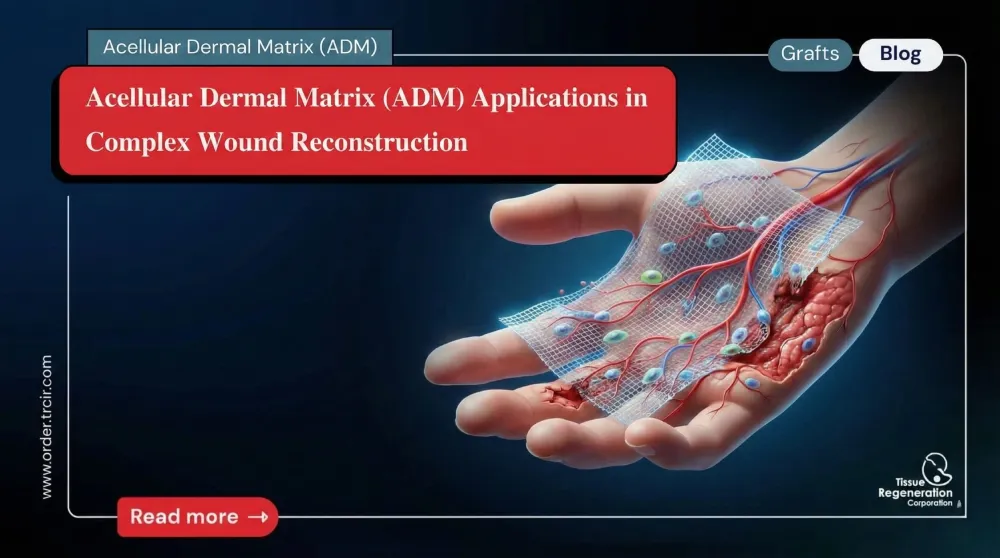
Before diving into the specific applications, it is essential to understand what this material actually is. To define it simply, an Acellular Dermal Matrix (ADM) is a soft tissue graft that functions as a biological scaffold.
It is derived from natural tissue—typically human (allogenic) or animal (xenogenic, usually porcine or bovine) skin. Through a sophisticated processing method known as decellularization, all genetic material, cells, and antigens that could cause an immune rejection are removed. What remains is a pure, sterile structural framework composed of collagen, elastin, and proteoglycans.
Think of it as the frame of a house. The frame itself doesn’t have residents yet, but it provides the necessary structure for people (in this case, the patient’s own cells) to move in and build a home. When ADM in complex wound reconstruction is utilized, it serves as a template that guides the body’s repair mechanism, allowing for the regeneration of healthy tissue rather than the formation of scar tissue.
How Does ADM Work in Wound Healing?
You might be wondering how a piece of decellularized tissue can actively help close a complex wound. The mechanism is quite fascinating and relies on the body’s natural biology.
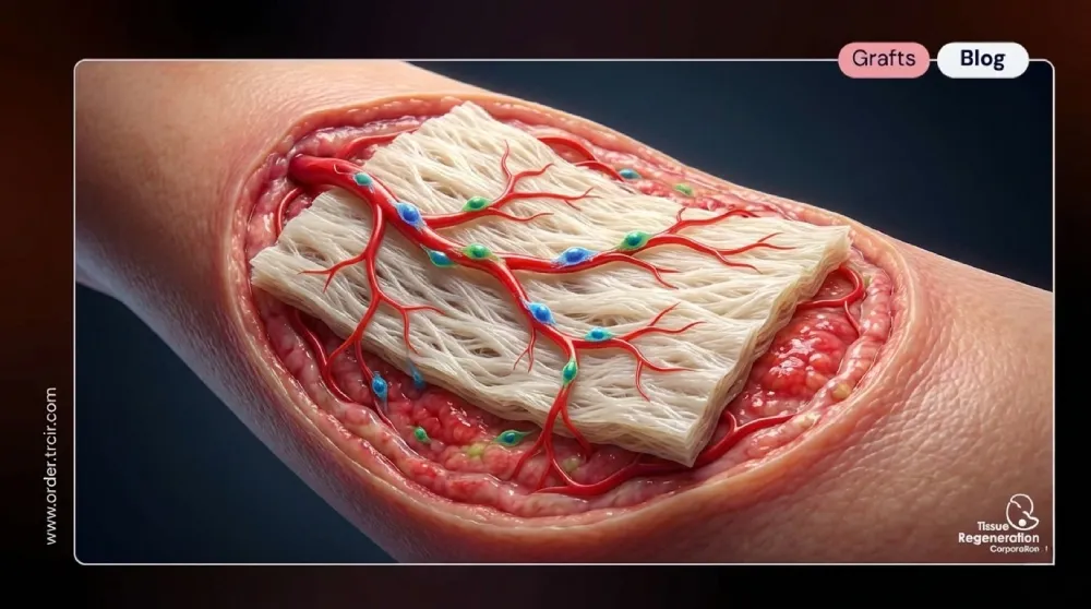
When the ADM is placed over a wound bed, it doesn’t just sit there like a bandage. It begins a biological interaction with the host tissue. Here is the step-by-step process of how it aids reconstruction:
- Scaffold Integration: The ADM provides a 3D structure that mimics the human extracellular matrix.
- Revascularization: The patient’s body begins to grow new blood vessels (angiogenesis) into the matrix. This is crucial because blood brings oxygen and nutrients needed for healing.
- Cellular Repopulation: Fibroblasts and other regenerative cells migrate into the ADM scaffold.
- Remodeling: Over time, the body slowly degrades the ADM material and replaces it with its own new, healthy collagen.
This process transforms the graft from a foreign material into the patient’s own living tissue, making ADM in complex wound reconstruction a permanent solution rather than a temporary fix.
Key Applications of ADM in Complex Wound Reconstruction
Now that we understand the “what” and “how,” let’s explore the “where.” ADM is versatile and is used across various surgical disciplines. Here, we will review the most critical applications where this technology makes a significant difference.
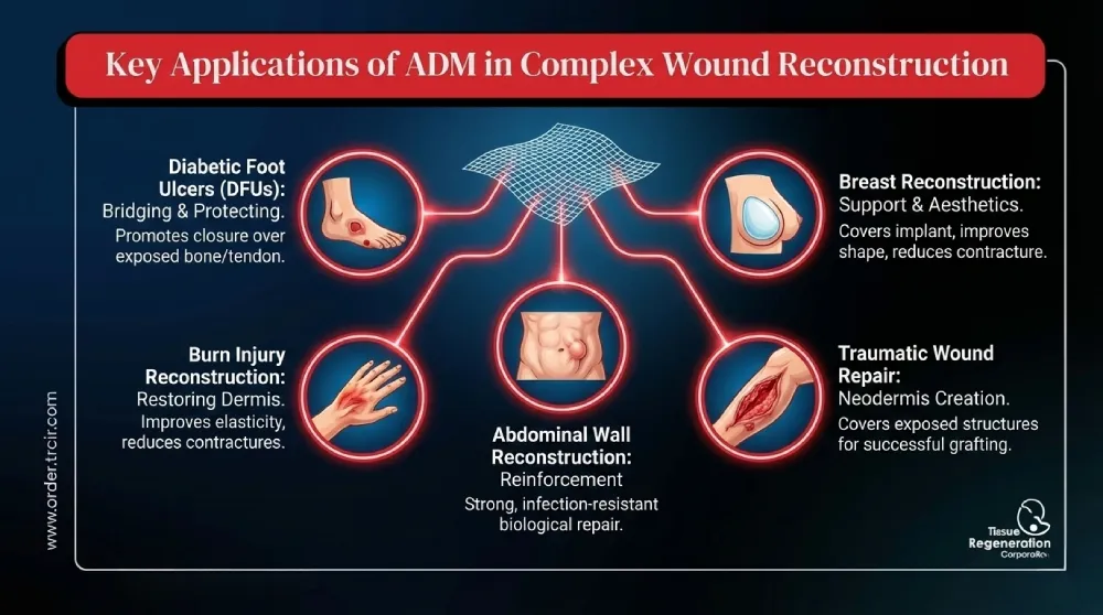
- Management of Diabetic Foot Ulcers (DFUs)
One of the most challenging conditions in wound care is the diabetic foot ulcer. Due to poor circulation and neuropathy, these wounds often refuse to heal and can lead to amputation.
ADM in complex wound reconstruction has shown remarkable success in this area. By applying ADM to a clean, debrided ulcer, clinicians provide a bridge for skin to grow over bone and tendon exposure. It protects vital structures and accelerates the closure rate significantly compared to standard moist wound therapy. For clinics sourcing supplies from order.trcir.com, high-quality ADM options can be a game-changer for limb salvage.
- Burn Injury Reconstruction
Deep partial-thickness and full-thickness burns destroy the dermis, the layer of skin responsible for elasticity and strength. Traditional skin grafts often result in tight, contracted scars because the dermis is missing.
Using ADM in burn care helps replace that missing dermal layer. It improves the pliability of the healed skin, reduces contractures (tightening that limits movement), and results in a better cosmetic appearance. It effectively reconstructs the skin’s architecture, not just its surface.
- Abdominal Wall Reconstruction (Hernia Repair)
In cases of complex ventral hernias or trauma where there is insufficient muscle or fascia to close the abdomen, surgeons need a strong reinforcement material. Synthetic meshes are often used, but they carry risks of infection and erosion into the bowel.
ADM is widely used here because it is resistant to infection and creates a strong, biological repair. It reinforces the abdominal wall and integrates with the muscles, providing a durable solution for patients who cannot tolerate synthetic implants.
- Breast Reconstruction
Perhaps the most common use of ADM today is in post-mastectomy breast reconstruction. When a surgeon places a tissue expander or implant, they often need extra coverage to support the implant and define the breast pocket.
ADM in complex wound reconstruction allows surgeons to cover the lower pole of the implant, providing support like a hammock. This leads to better aesthetic outcomes, reduced capsular contracture rates, and the ability to perform single-stage reconstruction in some cases.
- Traumatic Wound Repair
Severe trauma, such as degloving injuries or road accidents, often leaves large defects with exposed bone, tendon, or hardware. Skin grafts usually fail in these environments because they need a vascular bed to survive.
ADM can be placed directly over these exposed structures to create a “neodermis.” Once the ADM is vascularized, a thin skin graft can be placed on top, ensuring successful coverage and salvage of the limb or tissue.
Advantages of Using ADM
Why should a surgeon choose ADM in complex wound reconstruction over other methods? The benefits are substantial and contribute to better patient outcomes.
- Reduced Donor Site Morbidity: Unlike autologous tissue flaps (taking tissue from another part of the patient’s body), ADM comes off the shelf. This means there is no second surgical site to heal, reducing pain and complications.
- Biocompatibility: Since the cellular components are removed, the risk of immune rejection is extremely low.
- Infection Resistance: Biological matrices generally handle bacterial colonization better than synthetic meshes, making them safer for contaminated wounds.
- Structural Support: ADM provides immediate structural integrity while promoting long-term regenerative healing.
- Cosmetic Improvement: By restoring dermal thickness, the final healed wound looks and feels more like natural skin, with better elasticity.
Preparation and Surgical Considerations
Successful use of ADM in complex wound reconstruction relies heavily on proper preparation. It is not a magic patch; it requires a disciplined surgical approach.
First, Debridement is critical. The wound bed must be free of necrotic (dead) tissue and infection. The ADM must be placed in contact with healthy, bleeding tissue to ensure it can absorb fluid and allow blood vessel ingrowth.
Second, Fixation is key. The matrix must be secured using sutures or staples to prevent shearing forces. If the matrix moves, the fragile new blood vessels cannot penetrate it, leading to graft failure.
Finally, Moisture Balance is essential. The graft needs to be kept moist but not overly wet. Specialized dressings, often available through medical suppliers like order.trcir.com, are used to maintain this delicate environment during the integration phase.
Potential Risks and Challenges
We have discussed the benefits, but a balanced view requires looking at the challenges. While ADM in complex wound reconstruction is highly effective, complications can occur.
- Seroma Formation: Fluid can accumulate under the matrix, preventing integration. Surgeons often use drains or meshing techniques to prevent this.
- Cost: ADM is generally more expensive than synthetic materials or simple dressings. However, the cost is often justified by the reduction in healing time and secondary surgeries.
- Integration Failure: In patients with severe comorbidities (like heavy smoking or uncontrolled diabetes), blood flow may be too poor for the ADM to vascularize, leading to graft loss.
The Future of Wound Care with ADM
The field of regenerative medicine is not standing still. Research is continuously improving the processing methods of ADMs to retain more natural growth factors and structural proteins.
We are seeing the development of “flowable” ADMs for irregular tunnel wounds and combination products that include antibiotics or silver to fight infection actively. As these technologies evolve, the ability to treat even the most catastrophic wounds will improve, offering hope to patients who previously had few options. At order.trcir.com, we are committed to staying at the forefront of these medical advancements.
Frequently Asked Questions (FAQ)
To further clarify the topic of ADM in complex wound reconstruction, we have compiled some of the most frequently asked questions.
Is the use of ADM considered a permanent solution?
Yes. Unlike a synthetic mesh that remains as a foreign body forever, ADM is gradually remodeled by your body. Over time, it is replaced by your own collagen and cells, effectively becoming a natural part of your tissue.
How long does it take for ADM to heal?
The integration process typically begins within the first week. Vascularization (blood vessel growth) usually occurs within 2 to 3 weeks. However, full maturation and remodeling of the tissue can take several months, depending on the patient’s overall health and the size of the wound.
Is there a risk of disease transmission from ADM?
The risk is exceedingly low. The manufacturing process involves rigorous sterilization and decellularization that removes viruses and bacteria. Regulatory bodies have strict standards for tissue banking and processing to ensure patient safety.
Can ADM be used on infected wounds?
While ADM is more resistant to infection than synthetic mesh, it is generally recommended to clear active infections before implantation. However, some specific types of ADM are designed to be used in contaminated fields, provided that aggressive debridement and antibiotic therapy are also utilized.
Does insurance cover ADM reconstruction?
In many cases, yes. Insurance providers typically cover ADM in complex wound reconstruction when it is deemed medically necessary, such as for breast reconstruction post-mastectomy, severe burns, or chronic non-healing ulcers that have failed standard treatments. It is always best to verify with specific providers.
Conclusion
In this article, we have thoroughly explored the role of ADM in complex wound reconstruction. We discussed what this biological material is, how it acts as a scaffold for the body’s natural healing processes, and the wide array of clinical applications—from diabetic ulcers to complex hernias.
We also examined the significant advantages, such as reduced scarring and lack of donor site pain, while acknowledging the necessity of proper surgical technique to avoid complications.
It is clear that Acellular Dermal Matrix represents a monumental shift in how we approach tissue repair. It allows the body to regenerate rather than just repair, restoring form and function in ways previously thought impossible.
We hope this article has helped you gain a better understanding of this advanced medical technology. Whether you are a provider seeking the best tools for your patients or an individual researching your own care, knowledge is the first step toward recovery. For access to top-tier medical supplies and further information, remember to visit order.trcir.com.
References
- Orgill, D. P., & Ogawa, R. (2013). Acellular dermal matrix in reconstruction procedures. Plastic and Reconstructive Surgery.
- Brigido, S. A. (2006). The use of an acellular dermal regenerative tissue matrix in the treatment of lower extremity wounds: a prospective retrospective study. International Wound Journal.
- Nahabedian, M. Y. (2010). Allocating the use of acellular dermal matrices in breast reconstruction. The Breast Journal.
- Sanders, L., & Landsman, A. (2011). Surgical Management of the Diabetic Foot using Acellular Dermal Matrix. Clinics in Podiatric Medicine and Surgery.
- Janis, J. E., et al. (2012). use of Acellular Dermal Matrix in Abdominal Wall Reconstruction. Hernia.