
Dental medicine has undergone a revolution in the last few decades. The ability to replace missing teeth with dental implants has restored function and confidence to millions. However, the success of implants—and many other dental procedures—hinges on one critical factor: the presence of sufficient, healthy jawbone. When bone is lost due to trauma, extraction, or disease, the challenge for clinicians is to not just stop the loss, but to rebuild what’s been lost. This is the field of dental bone regeneration.
But how do we ensure this new bone is strong, stable, and integrates perfectly? The question of how to improve bone regeneration in dentistry is at the forefront of dental research. It’s a complex process that involves balancing advanced materials, surgical skill, and the body’s own healing capabilities. At trcir.com, we believe in empowering patients through education, and this guide explores the sophisticated techniques used in modern dentistry to achieve predictable and successful bone growth.
The Critical Need for Bone in Modern Dentistry
Bone is a dynamic, living tissue. In the jaw, the alveolar bone exists to support the teeth. When a tooth ittts lost, the underlying bony tissue no longer receives the functional stimulation it needs and begins to resorb, or shrink. This atrophy can happen remarkably quickly, leading to several complications:
- Dental Implant Instability: Implants require a solid foundation of bone to achieve osseointegration (the direct fusion of bone to the implant). Insufficient bone volume is the number one contraindication for implant placement.
- Periodontal Disease: Advanced gum disease (periodontitis) actively destroys the bone supporting the teeth, leading to mobility and eventual tooth loss. Regeneration is needed to save these teeth.
- Socket Preservation: After a tooth extraction, the empty socket will heal, but often with significant bone loss. Bone regeneration procedures at the time of extraction can preserve the ridge’s shape and volume, making a future implant much simpler.
- Sinus Augmentation: In the upper jaw, the maxillary sinuses (empty air-filled spaces) can sit very close to the roots of the back teeth. When these teeth are lost, there is often only a thin wall of bone remaining. A “sinus lift” or augmentation procedure is required to add bone into the sinus floor to create enough height for an implant.
Understanding the Gold Standard: Guided Bone Regeneration (GBR)
For decades, the foundational technique for how to improve bone regeneration in dentistry has been Guided Bone Regeneration (GBR). The concept is ingeniously simple and based on a biological principle: different tissues in the body heal at different speeds.
After an extraction or injury, the soft, fleshy gum tissue (gingival epithelium) grows much faster than new bone. Without intervention, this fast-growing soft tissue will rush into the empty space, filling it before the slower-moving bone cells get a chance. This results in a healed site that looks closed but is filled with soft tissue, not the strong bone needed for an implant.
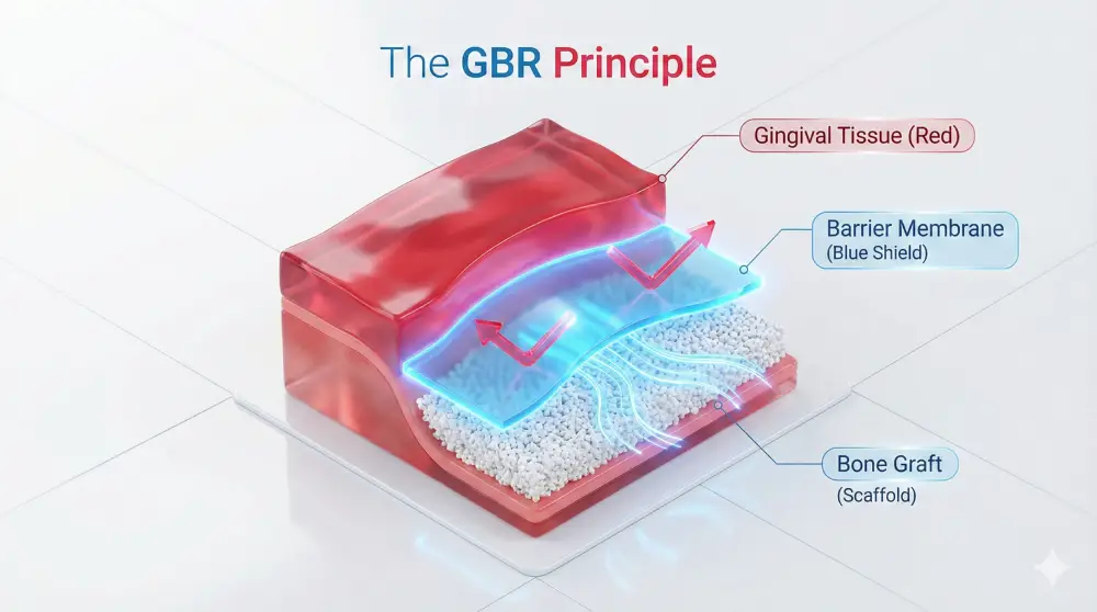
GBR solves this problem by using a barrier membrane. This is a small, thin sheet (like a tiny, sophisticated tent) that is placed over the bone graft material and under the gums.
This membrane serves two critical purposes:
- Exclusion: It physically blocks the fast-growing soft tissue cells from invading the defect.
- Space Maintenance: It creates and holds a protected, secluded space, allowing the void to be filled with a blood clot and graft material.
This protected space is the perfect environment for the body’s “construction crew”—slower-moving bone-forming cells (osteoblasts) and blood vessels—to migrate in, lay down a new bone matrix, and mature it into solid, functional bone.
The Materials That Make It Happen: Grafts and Membranes
A GBR procedure requires two key components: the “scaffold” (bone graft) to fill the space and the “shield” (membrane) to protect it. The choice of material is one of the most important decisions a clinician makes and is key to how to improve bone regeneration in dentistry for a specific patient’s needs.
Choosing the Right Bone Graft (The Scaffold)
The graft material acts as a scaffold, or an “osteoconductive” framework. It holds the space and provides a template for the new bone to grow upon. Some grafts are also “osteoinductive,” meaning they contain proteins that actively signal the body to create new bone.
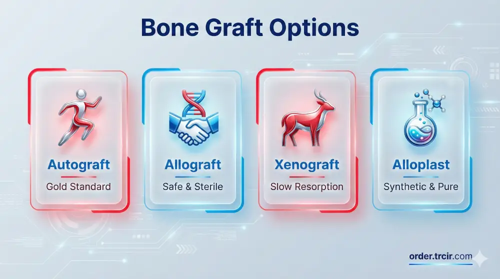
- Autografts: This is bone taken from the patient’s own body (e.g., from the chin, back of the jaw, or hip). It is the “gold standard” because it’s 100% compatible and osteoinductive (it has its own living cells and growth factors). The major drawback is the need for a second surgical site, which means more discomfort and potential complications for the patient.
- Allografts: These are grafts sourced from another human being. Human Tissue Allografts are harvested from deceased organ donors and processed through a stringent series of sterilization and screening procedures by accredited tissue banks. This process removes all cellular components, rendering the graft safe and non-immunogenic. It is a highly effective and very common choice, as it provides an excellent scaffold without the need for a second surgical site.
- Xenografts: This graft material is derived from an animal source, most commonly bovine (cow) or porcine (pig). The material is processed at very high temperatures to remove all organic matter, leaving only the mineral component of the bone. This inorganic scaffold is extremely similar to human bone, is very slow to resorb, and acts as an excellent space-maintainer for new bone to grow into.
- Alloplasts: These are synthetic, lab-made materials. They are typically composed of biocompatible minerals like hydroxyapatite (HA) or beta-tricalcium phosphate (β-TCP). They have the advantage of being completely sterile, risk-free, and available in unlimited supply. Their resorption rates can be engineered for different applications.
The Role of Barrier Membranes
The membrane is just as important as the graft.
- Resorbable Membranes: These are the most commonly used. They are typically made of collagen (sourced from bovine or porcine tissue) and are designed to last for several weeks or months—long enough for the initial bone growth to establish itself. After that, the body naturally and safely dissolves them, eliminating the need for a second surgery to remove them.
- Non-Resorbable Membranes: These membranes, made from materials like d-PTFE (Teflon) or reinforced with titanium mesh, provide a much more rigid and long-lasting barrier. They are exceptional for large defects where space maintenance is critical (e.g., building bone vertically). Their major disadvantage is that they must be removed in a second surgical procedure 6-9 months later.
Accelerating Healing: Biologics and Growth Factors
This is where the most exciting advancements are happening. If the graft is the scaffold and the membrane is the shield, biologics are the “super-fertilizer.” These are substances that concentrate the body’s own healing signals to supercharge the regenerative process. Using them is arguably the most effective method for how to improve bone regeneration in dentistry today.
Platelet-Rich Fibrin (PRF)
This is a revolutionary, 100% autologous (from the patient) biologic. The process is simple:
- A small amount of the patient’s blood is drawn, just like a standard blood test.
- The blood is immediately spun in a centrifuge using a specific protocol (with no anticoagulants).
- This separates the blood into layers. The clinician harvests the middle layer, a golden-colored clot of fibrin, which is packed with platelets, white blood cells, and a high concentration of growth factors (like PDGF, VEGF, and TGF-beta).
This PRF “clot” can be used as a membrane itself, or it can be chopped up and mixed with the bone graft material. When mixed with the graft, it creates what clinicians call “sticky bone”—a pliable, growth-factor-rich graft that is easy to handle, pack into the defect, and stays exactly where it’s placed. The fibrin matrix also releases these powerful growth factors slowly over 7-14 days, creating a sustained healing response.
Bone Morphogenetic Proteins (BMPs)
These are proteins that are powerfully osteoinductive. Their one and only job is to signal primitive stem cells in the area to turn into bone-forming osteoblasts. They don’t just help bone grow; they cause bone to grow.
The most common one used is BMP-2, a synthetic version of a natural human protein. Its Clinical Applications are typically reserved for very large defects, complex sinus lifts, or in patients with compromised healing. It is incredibly effective but is also significantly more expensive than other methods and carries its own specific set of protocols and potential side effects, such as post-operative swelling.
Beyond Materials: Surgical and Patient Factors
All the advanced materials in the world won’t work if the fundamentals aren’t right. How to improve bone regeneration in dentistry is as much about the patient and the procedure as it is about the products.
Surgical Technique
A successful GBR procedure is highly technique-sensitive. The surgeon must:
- Be Minimally Invasive: Preserve as much of the original tissue and blood supply as possible.
- Achieve Primary Closure: The gum tissue must be closed over the membrane and graft without any tension. If the flap is stretched too tight, the sutures will pull apart, the membrane will be exposed to the mouth, and the graft will become infected and fail.
- Ensure Stability: The graft and membrane must be immobilized. Any micro-movement will prevent blood vessels from growing in, leading to failure.
Patient Systemic Health
The patient’s overall health plays a massive role.
- Smoking: This is the #1 enemy of bone grafting. The nicotine and other toxins in cigarettes cause vasoconstriction (strangling of blood vessels), starving the healing site of the oxygen, nutrients, and cells it desperately needs. Most surgeons see a significantly higher failure rate in smokers.
- Uncontrolled Diabetes: High blood sugar impairs wound healing, reduces immune function, and increases the risk of infection.
- Nutrition: Healing requires protein, Vitamin C, Vitamin D, and other micronutrients. A poor diet can slow or stop the process.
- Post-Operative Compliance: The patient must follow instructions, including proper oral hygiene (e.g., antimicrobial rinses), a soft diet, and avoiding putting pressure on the area.
What’s Next? The Future of Dental Bone Regeneration
The quest for how to improve bone regeneration in dentistry is ongoing. The future is focused on even more precise, personalized solutions:
- Stem Cells: Harvesting mesenchymal stem cells (from the patient’s own bone marrow or fat) and introducing them to the site to provide a fresh supply of bone-forming cells.
- 3D-Printed Scaffolds: Creating custom-printed, perfectly-fitting scaffolds for a patient’s unique defect, possibly even pre-loaded with growth factors.
- Advanced Biologics: Moving beyond broad growth factors to using specific signaling pathways to control the speed and quality of bone formation.
Frequently Asked Questions About Bone Regeneration
What is the success rate of dental bone regeneration?
In a healthy, non-smoking patient, the success rate for routine bone grafting procedures (like socket preservation or GBR for implants) is very high, often cited at over 95%. Success depends on the size of the defect, the materials used, and patient compliance.
How long does it take for a dental bone graft to heal?
The graft material itself is just a scaffold. Your body needs time to resorb this scaffold and replace it with your own new, living bone. While the gums will heal in a few weeks, the bone maturation takes much longer, typically 4 to 9 months, depending on the size of the graft and the materials used. Only after this maturation period is the bone strong enough to receive a dental implant.
Is the dental bone regeneration procedure painful?
The procedure itself is not painful, as it is performed under local anesthesia, and you will be completely numb. Post-operative discomfort is expected and is very similar to that of a tooth extraction. This may include swelling, bruising, and soreness, which is typically well-managed with over-the-counter pain relievers (like ibuprofen) and ice packs.
Can my body reject a bone graft?
True “rejection” in the sense of an immune response is extremely rare. Processed Human Tissue Allografts and xenografts have all cellular and immunogenic components removed. The more accurate term is “failure.” A graft can fail if it becomes infected, if the membrane becomes prematurely exposed, or if the patient’s body cannot establish a good blood supply (e.g., in heavy smokers).
Partnering in Your Dental Health
How to improve bone regeneration in dentistry is a dynamic and evolving field. It has moved far beyond simply packing a hole with some material. Today, it is a sophisticated, multi-faceted approach that combines advanced materials like Human Tissue Allografts, biologic accelerators like PRF, and a deep understanding of the body’s healing processes.
The ultimate goal is to predictably and safely rebuild a living, functional bone foundation that can support dental implants for a lifetime. As we at trcir.com continue to follow these advancements, our focus remains on ensuring patients receive the most stable, aesthetic, and long-lasting solutions that modern dentistry can offer.
Scientific Sources
- Guided Bone Regeneration (GBR):
- Elgali, I., Omar, O., Dahlin, C., & Thomsen, P. (2017). Guided bone regeneration: A systematic review of the effect of barrier membranes on bone regeneration. Journal of Clinical Periodontology, 44(S18), O1-O15.
- Link: https://www.ncbi.nlm.nih.gov/pmc/articles/PMC5643444/
- Platelet-Rich Fibrin (PRF) in Dentistry:
- Miron, R. J., Fujioka-Kobayashi, M., Hernandez, M., et al. (2017). Platelet-Rich Fibrin (PRF): A Pervasive and Natural Fibrin-Based Biopattern for Tissue Regeneration. International Journal of Dental Sciences, 2017.
- Link: https://www.ncbi.nlm.nih.gov/pmc/articles/PMC5504780/
- Bone Morphogenetic Proteins (BMPs):
- Carter, T. G., & Brar, P. S. (2011). Bone morphogenetic proteins (BMPs) in periodontal and implant regeneration. Journal of the Indian Society of Periodontology, 15(1), 2.
- Link: https://www.ncbi.nlm.nih.gov/pmc/articles/PMC3134017/
- Bone Graft Materials Review:
- Amini, A. R., Laurencin, C. T., & Nukavarapu, S. P. (2012). Bone tissue engineering: recent advances and ongoing challenges. Journal of Materials Science: Materials in Medicine, 23(12), 2883–2899.
- Link: https://www.ncbi.nlm.nih.gov/pmc/articles/PMC3504938/