
The landscape of aortic valve replacement (AVR) has been profoundly reshaped over the last two decades. The rise of durable bioprosthetic xenografts and the revolutionary, minimally-invasive approach of Transcatheter Aortic Valve Replacement (TAVR) have captured the clinical spotlight, becoming the standard of care for the vast majority of patients with aortic stenosis.
In this new era, the aortic valve allogarft—a cryopreserved human aortic valve harvested from a deceased donor—can seem like a relic of a past generation. Its use has declined significantly, relegated to a niche status in most cardiac centers. This has led many to ask: Is there still a place for the allogarft?
The answer is a definitive and resounding yes.
While allogarfts are no longer the default choice for a routine AVR, they have settled into a critical role where they are not just an option, but the only option. For a specific, high-risk, and often desperate patient population, the aortic valve allogarft is not just relevant; it remains the undisputed gold standard. This article explores the modern role of the aortic valve allogarft, its unique advantages, its known limitations, and the specific clinical scenarios where it remains irreplaceable.
What is an Aortic Valve Allogarft?
An aortic valve allogarft (or allograft) is the complete aortic root—including the valve leaflets, the aortic annulus, and a portion of the ascending aorta—harvested from a human donor.
Unlike mechanical valves (made of carbon) or bioprosthetic xenografts (made from bovine or porcine tissue), the allogarft is a fully human biological entity. After retrieval, it undergoes a meticulous process of:
- Dissection and Sizing: The valve is carefully dissected from the donor’s heart and measured.
- Antibiotic Decontamination: It is treated with a cocktail of antibiotics to ensure sterility.
- Cryopreservation: The tissue is placed in a cryoprotectant solution and slowly frozen to ultra-low temperatures (typically -135°C or below) in liquid nitrogen vapor. This process, vital for tissue banking, arrests all biological activity and preserves the tissue’s structural integrity for years.
- Quality Control: Extensive serological testing is performed to screen for infectious diseases, ensuring the safety of the allograft.
Historically, the appeal of the allogarft was its near-perfect central-flow hemodynamics (mimicking a native valve) and its freedom from mandatory lifelong anticoagulation, a significant burden associated with mechanical valves. However, its long-term durability became its primary drawback.
The Challenge: Why Allogarfts Are Not for Everyone
The decline in allogarft use for routine AVR was driven by one primary factor: structural valve deterioration (SVD).
Despite being human tissue, the cryopreservation process renders the valve leaflets non-viable. The recipient’s immune system recognizes this non-living allograft tissue, triggering a slow, chronic inflammatory and immune response. Over time, this leads to leaflet stiffening, calcification, and eventual tearing, resulting in aortic stenosis or regurgitation.
This process is particularly aggressive in younger patients, whose more active immune systems can lead to allogarft failure in as few as 8-10 years. For a 50-year-old patient, this virtually guarantees at least one, if not more, complex re-operations in their lifetime.
Furthermore, implanting a allogarft is not a simple “drop-in” procedure. It is almost always a full aortic root replacement, a technically demanding operation that requires more time, skill, and experience than implanting a stented prosthetic valve.
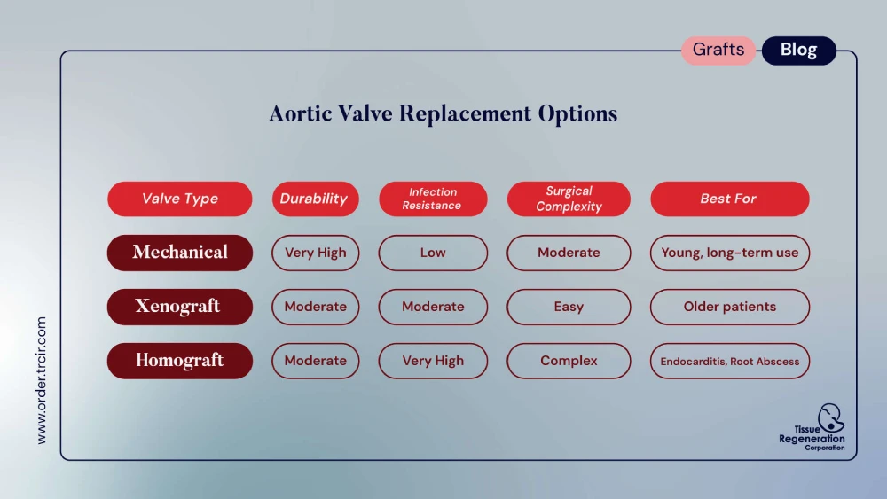
Given these challenges, the modern, readily available, and surgically simpler pericardial (bovine) xenografts—which offer a reliable 15-20 year lifespan in older patients and a pathway to future valve-in-valve TAVR—became the logical choice for most surgeons and patients.
The Gold Standard Indication: Active Infective Endocarditis
The allogarft’s story would end there, were it not for its unique, life-saving properties in the face of one of the most feared complications in cardiac surgery: active infective endocarditis (IE) with aortic root destruction.
Infective endocarditis is a bacterial or fungal infection of the valve structure. In its most destructive form, the infection is not contained to the leaflets but burrows deep into the aortic annulus, forming a large, pus-filled aortic root abscess.
This clinical scenario is a surgical nightmare for several reasons:
- Destroyed Anatomy: The abscess liquefies the tissue where a new valve must be sewn, leaving the surgeon with “nothing to sew to.”
- High Risk of Reinfection: Implanting a foreign body (like a mechanical valve or xenograft) into an actively infected field is extraordinarily risky. The prosthetic material provides a perfect surface for bacteria to colonize, leading to prosthetic valve endocarditis (PVE), which carries a mortality rate of over 50%.
This is precisely where the aortic valve allogarft is unparalleled.
1. Unmatched Resistance to Infection
The allogarft’s greatest strength is biological. As a devitalized human tissue, it is remarkably resistant to recurrent infection. Unlike the fabric sewing ring of a prosthetic valve—a prime target for bacterial biofilm—the allogarft tissue seems to manage and clear residual bacterial load in a way no synthetic material can. Numerous clinical studies have confirmed that the incidence of recurrent endocarditis is significantly lower with allogarfts compared to any other valve substitute in this setting.
2. The Ultimate Surgical “Patch”
The second, and perhaps more important, advantage is surgical versatility. An aortic root abscess requires radical debridement—the surgeon must cut away all infected, necrotic tissue, often leaving a massive, irregular defect in the heart.
A stented prosthetic valve is a fixed, rigid device. It cannot be used to “patch” this defect.
The aortic root allogarft, however, is a large, pliable, and tailorable piece of biological tissue. It comes with the aortic root, the valve, and often the anterior leaflet of the mitral valve attached. This gives the surgeon a complete “biological toolkit” to:
- Replace the entire aortic root.
- Reconstruct the left ventricular outflow tract (LVOT).
- Patch defects in the aorto-mitral curtain.
- Re-implant the coronary arteries.
In short, the surgeon can radically excise the entire abscess and then use the allogarft to rebuild the heart’s anatomy from scratch. This procedure is simply not possible with any other valve substitute. For this reason, in guidelines from the American Heart Association (AHA) and the European Society of Cardiology (ESC), the aortic valve allogarft is consistently named as the device of choice for AVR in the setting of destructive endocarditis with root abscess.
Other Key Applications for the Allogarft
While endocarditis is its primary “gold standard” indication, the allogarft remains a vital tool in other complex scenarios.
The Ross Procedure: The Pulmonary Autograft
The Ross procedure is an elegant operation performed in children and young adults (typically under 40) with aortic valve disease.
- The patient’s diseased aortic valve is removed.
- The patient’s healthy pulmonary valve (the autograft) is harvested and moved into the aortic position.
- This autograft is a living valve. It does not degenerate, does not require anticoagulation, and grows with the patient, making it the ideal lifetime aortic valve replacement.
This, however, leaves a defect in the pulmonary position. A pulmonary valve allogarft is the overwhelming choice to replace the harvested pulmonary valve. In the low-pressure, right-sided pulmonary system, the immune-mediated SVD that plagues aortic allogarfts is dramatically slowed. A pulmonary allogarft can last 20 years or more, making it the perfect companion to the Ross procedure.
Complex Re-Operations and Congenital Heart Disease
The allogarft’s surgical flexibility also makes it a “bail-out” device for highly complex re-operations. In patients with multiple previous sternotomies, distorted anatomy, or complex congenital conditions (like truncus arteriosus), the allogarft provides a conduit that can be tailored to fit abnormal anatomy in a way that a rigid prosthetic cannot.
The Future: Decellularized Allogarfts
The primary limitation of allogarfts has always been durability. But what if you could have the perfect structure and versatility of a allogarft without the immune-mediated SVD?
This is the goal of next-generation tissue engineering. New techniques are emerging to create decellularized aortic valve allogarfts.
Using detergents and enzymatic solutions, the donor cells (which trigger the immune response) are completely washed away, leaving behind a pristine, acellular scaffold of collagen and elastin (the dECM, or decellularized extracellular matrix). The idea is that this “blank” scaffold, when implanted, will not be attacked by the immune system. Instead, it will be slowly repopulated by the patient’s own cells, potentially transforming it into a living, durable, and non-immunogenic valve.
While still in clinical investigation, these advanced allogarfts represent the next frontier, merging the best of allograft biology with the principles of regenerative medicine.
The Verdict: A Niche, But a “Golden” One
So, is the aortic valve allogarft still a gold standard?
- For routine aortic valve replacement? Its limited durability and surgical complexity mean it has been rightly surpassed by xenografts, mechanical valves, and TAVR.
- For the right patient?
The allogarft has evolved from a generalist tool to a high-precision instrument for the most complex and life-threatening cases. For the patient with a destructive aortic root abscess from endocarditis, the aortic valve allogarft is not just “an” option—it is the gold standard, the benchmark against which all other therapies are measured. It offers the best chance of survival by simultaneously solving two problems: eradicating infection and reconstructing the heart.
In an age of high-tech devices, the humble human allogarft remains one of the most powerful and irreplaceable tools in the cardiac surgeon’s armamentarium.
Sources and Further Reading
- American Heart Association (AHA) / American College of Cardiology (ACC) Guidelines for the Management of Valvular Heart Disease:
- Provides the official clinical recommendations, often citing allogarfts for complex endocarditis.
- https://www.ahajournals.org/journal/circ (Search for “2020 Guideline for the Management of Valvular Heart Disease”)
- European Society of Cardiology (ESC) Guidelines on Endocarditis (2023):
- The latest European guidelines detailing the surgical management of IE, including the role of allogarfts.
- [https://www.escardio.org/Guidelines/Clinical-Practice-Guidelines/Infective-Endocarditis]
- Jahanyar, J., & M.D. F, et al. (2020). “Aortic root replacement with allogarft for prosthetic valve endocarditis: A single-center experience.” Journal of Cardiac Surgery.
- A clinical study demonstrating the excellent outcomes of using allogarfts for the complex problem of prosthetic valve endocarditis.
- [https://onlinelibrary.wiley.com/doi/10.1111/jocs.14930]
- Arabkhani, B., et al. (2016). “Allogarft aortic root replacement in native and prosthetic active infective endocarditis: a review of the literature.” Annals of Cardiothoracic Surgery.
- A comprehensive review article that supports the allogarft’s role as the “gold standard” in endocarditis with root destruction.
- [https://www.ncbi.nlm.nih.gov/pmc/articles/PMC5075841/]
- David, T. E. (2018). “Aortic Valve-Sparing Operations and Aortic Root Replacement.” Surgical Clinics of North America.
- Discusses the technical aspects of aortic root surgery, highlighting the versatility of different conduits, including allogarfts.
- [https://www.surgical.theclinics.com/article/S0039-6109(18)30064-0/fulltext]